


DI PLASTIC SURGERY
DI DORSAL-HUMP RHINOPLASTY
What is dorsal-hump rhinoplasty?
A nasal hump can result from abnormal nasal-bone growth or post-traumatic changes such as fractures, creating a raised area in the middle of the nasal bridge. DI Dorsal-Hump Rhinoplasty smooths the protruding bridge to create a softer impression.
Surgery information
- Surgery duration
- 1 hour 30 minutes
- Anesthesia
- Sedation / general anesthesia
- Hospital admission
- Not usually required
- Recovery period
- Varies by individual
- Suture removal
- Required
- Follow-up visits
- 2–3 visits
Bleeding, infection, inflammation, bruising, swelling and pain may occur. Satisfaction and outcomes vary between individuals.

DI PLASTIC SURGERY
THROUGH A DETAILED ASSESSMENT
Through precise assessment
A considered approach to a balanced
Nasal contour!
Although it may appear simple from the outside, The inside of the nose has a complex structure. DI Plastic Surgery considers the overall harmony and appearance of the face and nose, 3D CT equipment is used for detailed assessment.
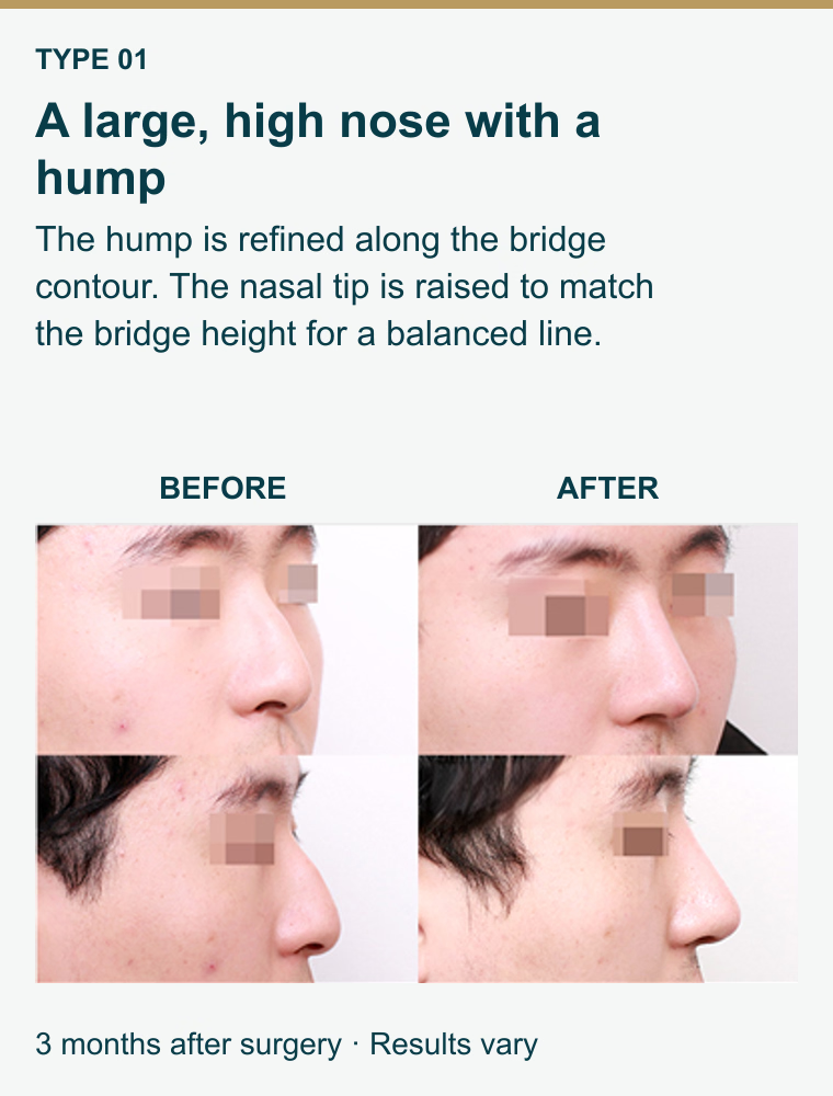
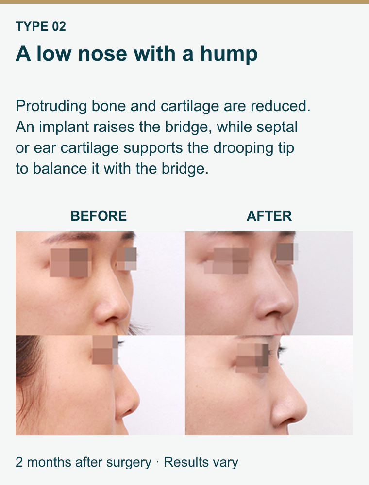
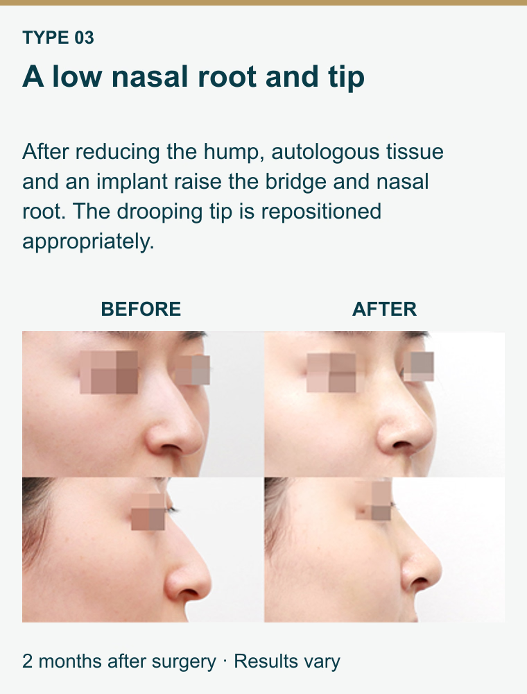
Dorsal hump correction: tailored to your nose
DI PLASTIC SURGERY
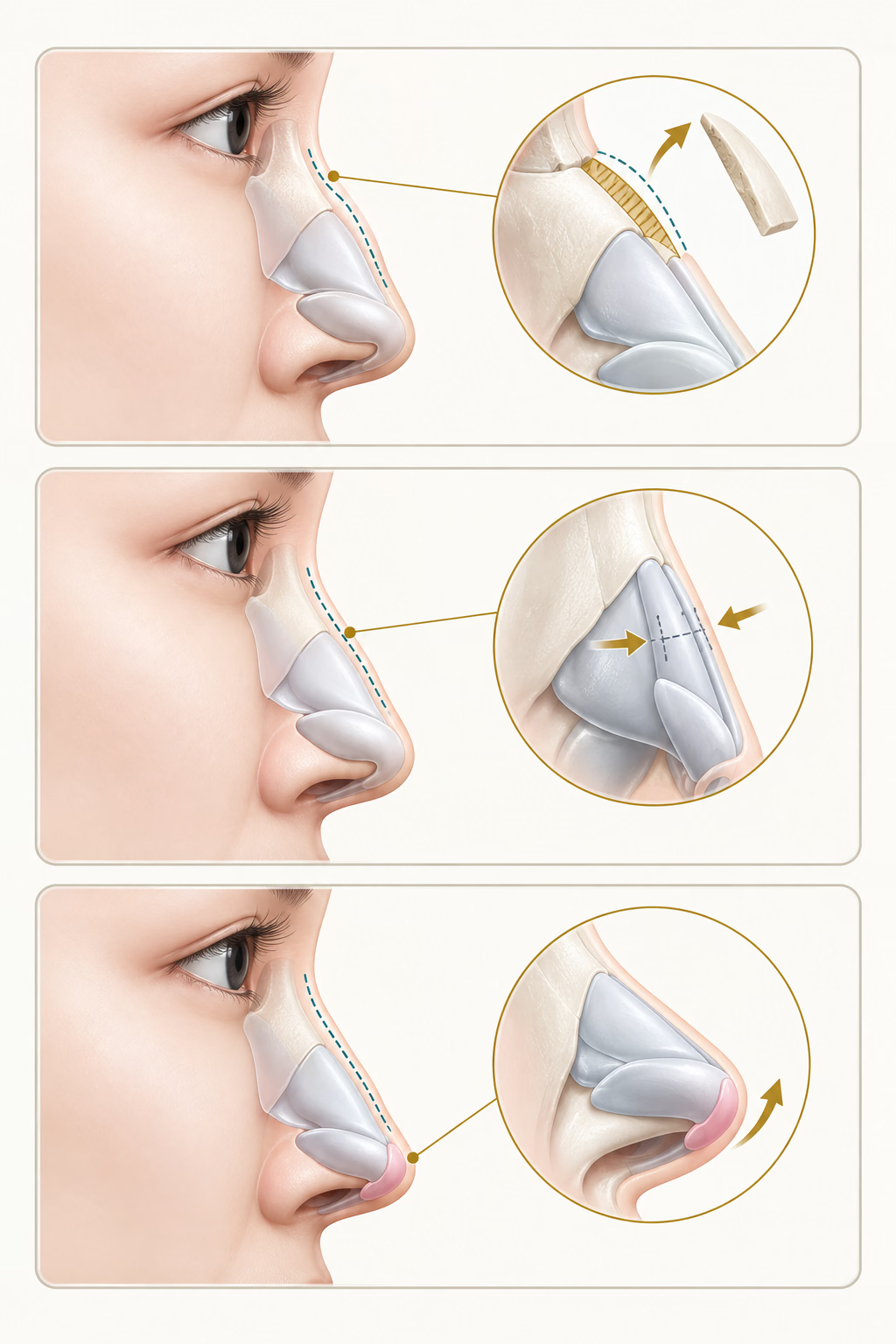
DI Dorsal-Hump Rhinoplasty: Surgical Method
Solution 01
Nasal hump with a low nasal root
The bridge prominence is removed and the nasal root-to-tip contour is adjusted as a whole.
Remove the protruding hump; if the bridge is low, insert an implant
Solution 02
Nasal hump with a high nasal root
Protruding bone and cartilage are reduced to create a smooth, even surface.
At the protruding hump, bone and cartilage are smoothed
Solution 03
A high nasal root with a drooping tip
and nasal hump
The hump is removed and the nasal tip is adjusted to harmonize with the bridge.
Protruding hump removal
Tip design using the patient's own cartilage
DI PLASTIC SURGERY
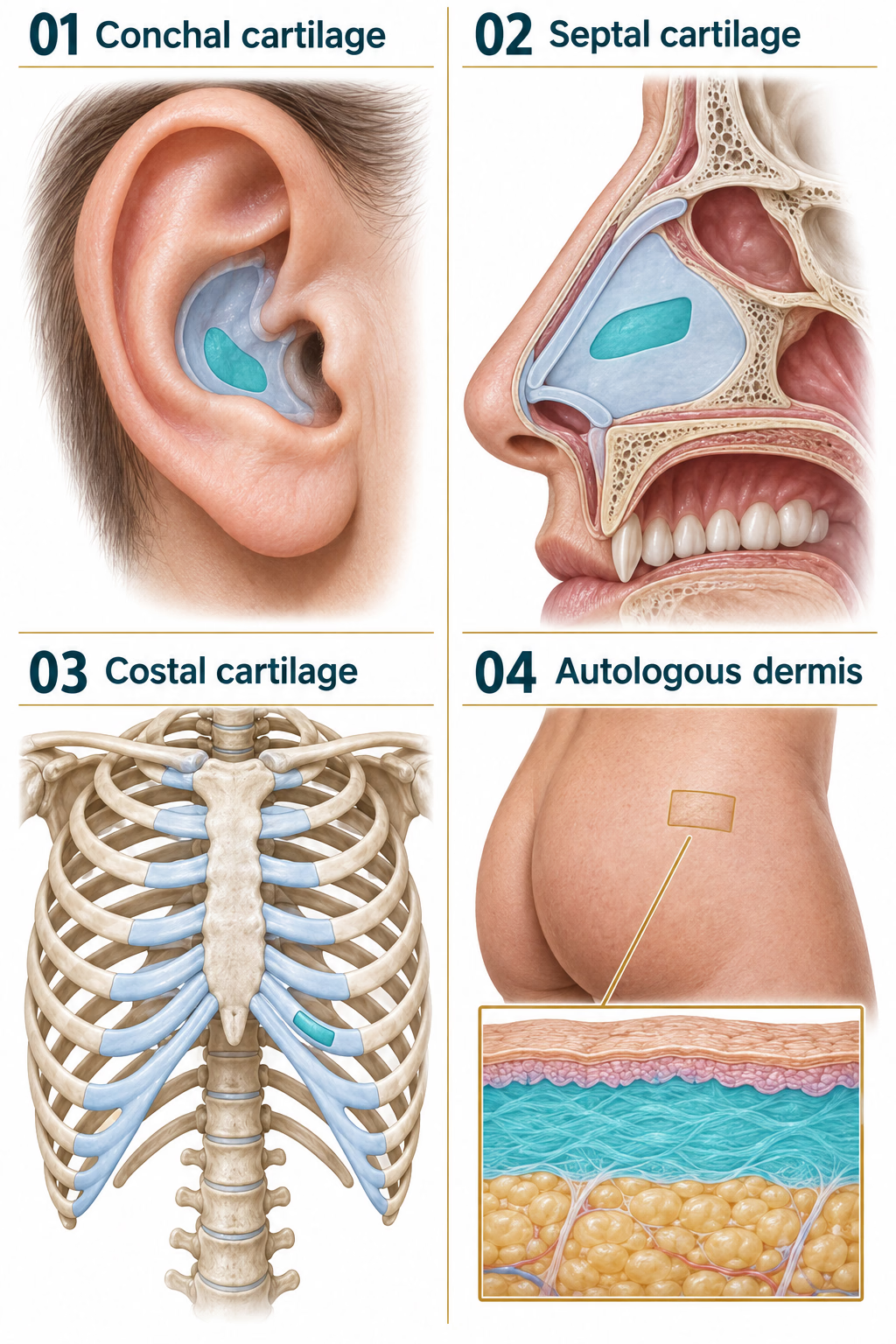
Features of DI autologous-tissue rhinoplasty
01. Ear cartilage
Cartilage from the conchal bowl of the ear can support and refine nasal-tip contours. The incision is placed discreetly, and the remaining ear structure is preserved.
02. Septal cartilage
Firm, plate-like cartilage from the internal nasal septum can support the nasal tip and other structures. A supporting border of septal cartilage is retained.
03. Rib cartilage
Costal cartilage from the anterior chest may be used when other available cartilage is insufficient. It provides material and structural support for the tip, bridge and other applications.
04. Autologous dermis
Dermis is a skin layer, not cartilage. A dermal graft may be used to add coverage or volume when skin is thin or when an implant is not preferred.
Illustrated donor areas are a general guide. The exact site and amount of tissue are selected for each individual.
Suited to my face
For your nasal contour, choose DI!
Straight profile
A straight line when viewed from the side
A profile often requested by men
Gently curved profile
A slight curve compared with a straight profile
A contour commonly preferred by women
Curved profile
A more pronounced curve
Upturned profile
An elevated tip with a low bridge
DI PLASTIC SURGERY
DORSAL-HUMP RHINOPLASTY
Who may consider this procedure?
People with a raised mid-bridge area and an uneven nasal contour
People seeking a smooth nasal bridge
People concerned about a strong or sharp impression
People with a high bridge but an irregular nasal shape

DI PLASTIC SURGERY
DORSAL-HUMP RHINOPLASTY
Why consider DI?
Individual facial form and characteristics guide surgery tailored to the nasal hump. Surgery is performed according to this plan.
3D-CT is used to examine nasal structures.
Nasal cartilage and internal structure are accurately assessed before surgery.
A thorough aftercare system supports you after surgery.

DI PLASTIC SURGERY
CHOSEN BY DI PATIENTS
Using your own cartilage for nasal tip surgery
New-material implants
Artificial implants
Synthetic cartilage materials that may occur inflammation and other adverse effects
Using the patient's own cartilage More natural movement
Integrated autologous cartilage Semi-permanent retention

Rhinoplasty aftercare
Follow your surgical team’s individual instructions. Recovery and follow-up schedules vary.
- 01
Incision care
Keep the surgical area dry and clean until stitches are removed. On the day of removal, wash gently with water; use soap from the following day.
- 02
Nasal discharge
A little bleeding or blood-tinged discharge may occur for 3–4 days. Do not blow your nose; gently wipe away discharge.
- 03
Tape and splint
Tape helps maintain implant position and limit blood pooling. The splint protects against impact. If it comes off, do not reattach it yourself; contact the clinic and protect your nose.
- 04
Cold and warm compresses
Use cold compresses for the first 3 days, then warm compresses from day 4, as instructed by your surgical team.
- 05
Swelling and bruising
Swelling and bruising generally take about 2 weeks to settle. Swelling may temporarily make the nose look uneven, tilted or depressed.
- 06
Glasses
Avoid glasses and sunglasses for at least one month to prevent pressure that could affect the nasal shape.
- 07
Protect your nose
Do not blow your nose forcefully or do activities that strain or put pressure on your nose.
- 08
Ear cartilage donor site
If cartilage was taken from your ear, avoid pulling or pressing the ear firmly.
- 09
Recovery habits
Avoid alcohol and smoking for one month. Avoid strenuous exercise and excessive stress, which can interfere with recovery.